Musee d’Anatomie Delmas-Orfila-Rouviere


France | Paris

Website 

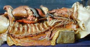
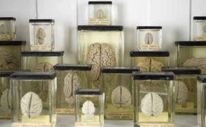
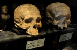
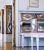
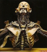
About

France’s largest collection of anatomical models and specimens began as small collection cultivated by Honore Fragonard (a professor of anatomy at the University of Paris). The collection was enhanced by Mathieu Orfila ( the dean of Medicine at U. of Paris), after he visited London’s Huntarian. In true french patriotism he sought to out shine the British. The museum contains specimens of humans and animals, including embryological reconstructions and pieces of neuro-anatomy.

Relevant People:

Honore Fragonard

Musée d’Anatomie de Montpellier


France | Montpellier

Website 

About

The oldest and the largest anatomy museum in France, the museum is situated inside the world famous and historic University of Montpellier. Structured with wide Doric columns, the museum‘s collections were listed as historic monuments in 2004. The museum’s educational dimension cannot be missed amidst the eeriness which envelops it. The main features of the museum are: -Deformed foetuses preserved in jars -Human and animal skeletons -An “ethnological” collection of skulls.

Relevant People:

Kings College, London: The Gordon Museum of Pathology


England | London

Website 

About

Gordon museum is the largest medical museum in the United Kingdom located in Kings College, London. Their collection includes many historically important artifacts including the Joseph Towne anatomical and dermatological wax models, the Lam Qua pre-operative tumour paintings and the specimen and artefact collections of Hodgkin, Thomas Addison, Bright and Astley Cooper.

Relevant People:

University College London: Grant Museum of Zoology


England | London

Website 

About

Founded in 1828, the Grant Museum houses over 67,000 specimens, covering the entire Animal Kingdom. Their collections include some rare and even extinct animal skeletons such as the Tasmanian Tiger or Thylacine, the Quagga, and the Dodo. The Brain Collection Most of the specimens were originally brought from the Gordon museum. Each brain is preserved in alcohol and suspended in glass jars. The collection is made up entirely of mammals, except for a single turtle, allowing the visitors a comparative tour of mammalian brains. The brains were collected from Africa, Asia, South America and Australia and Europe. Most of the brains are whole, a few have been dissected to show the internal anatomy. Of particular interest are the tiger cub that was only a few hours old, and the domestic dog. The dog’s brain has been annotated and each section of the brain sports a tiny label that has been stapled to it.

Relevant People:

Croatian Institute for Brain Research


Croatia | Zagreb

Website 

About

The Croatian Institute for Brain research is an affiliate of the University of Zagreb Medical School, and an independent research facility and archive. Its founder Dr. Ivica Kostovic established Zagreb’s neuroembryological collection; a significant collection of preserved human brains at various stages of development. It is one of the largest collections of human brains in the world consisting of 1,300 developing and adult brains. It has served as a valuable resource for many research projects, more than 30 PhD theses and upwards of 200 publications. The Institute even has a dormitory for researchers wishing to visit and utilize the collection. However, the institute is currently creating an online catalogue of the collection that will be readily available to those who cannot travel all the way to Croatia.

Relevant People:

Professor Ivica Kostovic

Josephinum Collections of the Medical University of Vienna


Austria | Vienna

Website 

About

The famous collection of wax anatomical and obstetric models is the result of a personal initiative of Joseph II. Inspired by the wax models in “La Specola” in Florence, commissioned by his brother (the Grand Duke of Tuscany and later Emperor Leopold II), Joseph II ordered 1,192 models for the newly-founded academy in Vienna. These were produced in Florence between 1784 and 1788 under the supervision of director Felice Fontana and anatomist Paolo Mascagni and eventually arrived in Vienna after a wearisome journey over the Alps. They were intended to serve on the one hand as visual aids for teaching, but on the other they were, even then, intended for the general public. The majority of the models survived to this day and they are exhibited in six rooms in their original rosewood and Venetian glass display cases.

Relevant People:

The Museum of Anatomy in Innsbruck


Austria | Innsbruck

Website 

About

The university of Innsbruck, founded by the emperor Leopold I, started its first lectures in 1672. The first anatomical professorship was given to Prof. Theodor Friedrich von STADTLENDER on April 22th 1689. The most important instrument for instructions in anatomy was then, as it is today, the human body. The „Letztwillige Verfügung“, i.e. the disposition of one’s body by will, did not exist in the 17th century. Therefore it was quite a problem to procure corpses and opportunities for dissection, the more so since the dissecting of people was thought to be a sin by many people. One possibility to get suitable corpses for dissection, however, there was: the anatomy professor could try to acquire from the hangman the dead bodies of persons who had been sentenced to death (death penalty being the customary punishment then for many crimes).

Relevant People:

Brain Museum of the Jacobs School of Medicine and Biomedical Sciences


United States | Buffalo

Website 

Brain Museum

About

Taken as a whole, the Brain Museum’s collection demonstrates how the components of our nervous systems work together. Visitors can see the brain’s layers and internal structures firsthand and view them from different angles.

At the original South Campus location, almost 90 beautifully illuminated brain specimens highlight anatomical features such as the corpus callosum, hippocampus and cerebellum. Pathological specimens show conditions such as Alzheimer’s disease, cerebral aneurysms and hydrocephalus.

Dissections show the full pathways for vision and hearing, and photographs offer closer views of the brain’s intricate structures. A display that highlights the achievements of groundbreaking researchers in neuroanatomy sets the discipline in its historical context.

The museum also houses a world-class collection of slides that display stained cross-sections of brain tissue, which medical students and researchers can consult by arrangement with the Department of Pathology and Anatomical Sciences.

Relevant People:

Harold Brody, MD

Second location at th Buffalo Niagara Medical Campus


Supported by
 
In collaboration with
FENS          IBRO EPFL          Experimental Museology Lab eM+
All information and images rights are reserved to the corresponding institutions. Theme and design: by David Martinez Moreno.